
Spinal disorders—ranging from cervical spondylosis and acute paraspinal muscle spasms to lumbar herniated discs and sciatica—represent the leading cause of global disability. In Vadodara, postural strain from sedentary desk work, industrial repetitive trauma, vehicular vibration, and progressive degenerative spine changes drive an increasing number of patients to seek specialized orthopedic evaluation.
Whether you are suffering from a sharp radiating pain down your leg, persistent neck stiffness limiting cervical rotation, or debilitating lower back spasms, understanding the biomechanics, underlying pathologies, and evidence-based interventions is the first step toward lasting functional recovery.
1. Anatomy and Biomechanics of the Spine
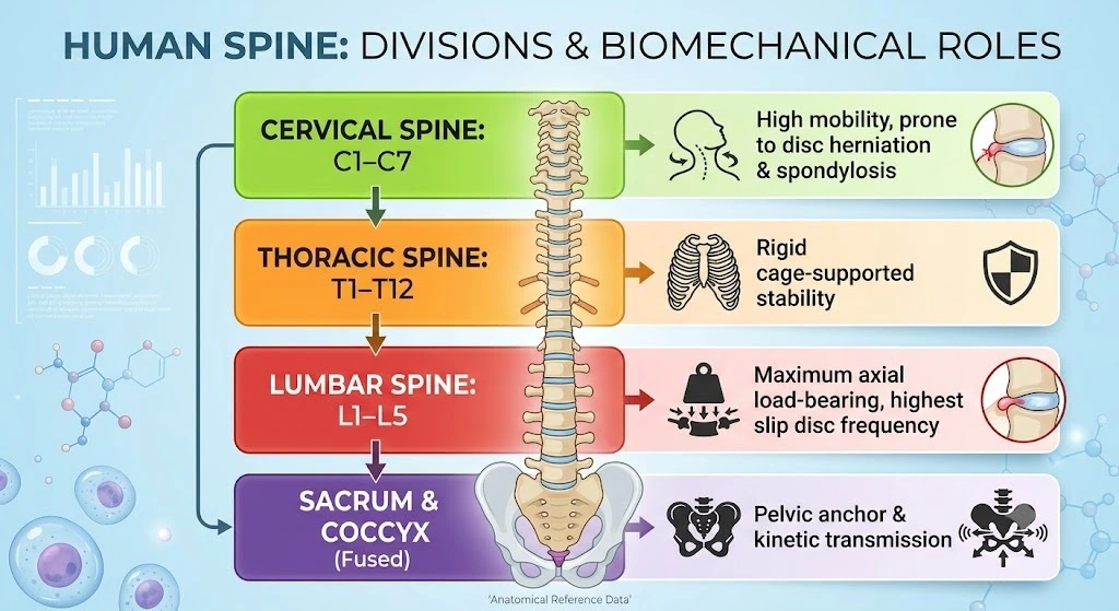
The human vertebral column consists of 33 vertebrae divided into five distinct anatomical regions: cervical (7), thoracic (12), lumbar (5), sacral (5, fused), and coccygeal (4, fused). It functions as the primary axial load-bearing pillar of the musculoskeletal system, protecting the central spinal cord and branching peripheral spinal nerves.
The Intervertebral Disc Complex
The intervertebral disc functions as a hydraulic shock absorber between adjacent vertebral bodies:
- Annulus Fibrosus: A robust outer ring composed of 15–25 concentric lamellae of Type I collagen fibers, oriented at alternating 60-degree angles to resist multidirectional torsion and shear forces.
- Nucleus Pulposus: A hydrated, gelatinous inner core containing rich concentrations of proteoglycans (predominantly aggrecan) and Type II collagen, maintaining high intradiscal oncotic pressure to absorb compressive axial stress.
- Cartilaginous Endplates: Thin hyaline cartilage layers interfacing the disc with the vascular vertebral body, governing nutrient diffusion into the essentially avascular disc space.
Cervical vs. Lumbar Biomechanical Dynamics
- Cervical Spine (C1–C7): Engineered for high ranges of motion across flexion, extension, lateral bending, and axial rotation. Due to this wide kinetic arc, C5–C6 and C6–C7 undergo rapid degenerative mechanical wear, resulting in cervical radiculopathy and disc osteophyte formation.
- Lumbar Spine (L1–L5): Built for significant axial compressive loading. The L4–L5 and L5–S1 segments bear the highest mechanical shear stress in the entire human body, making them the primary epicenter for disc protrusion, extrusion, and spinal stenosis.
2. Pathogenesis and Etiology: The Degenerative Cascade
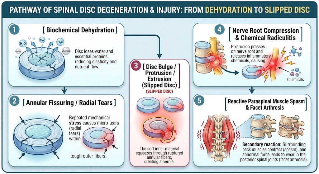
Spinal degeneration follows an orderly anatomical and pathological progression:
Slipped Disc Progression (Herniation Stages)
A “slipped disc” is a clinical lay term; the actual anatomical entity is a herniated nucleus pulposus (HNP). It develops across four defined stages:
- Disc Degeneration (Degenerative Disc Disease): Aging, microtrauma, and impaired endplate vascular diffusion deplete proteoglycans within the nucleus pulposus, causing dehydration and mechanical height loss.
- Disc Prolapse / Bulge: The nucleus displaces posteriorly or posterolaterally into an intact or weakened inner annulus, creating a smooth, symmetrical, or broad-based bulge into the spinal canal.
- Disc Extrusion: Complete rupture of the annulus fibrosus allows the soft, nuclear gel to breach the outer boundaries of the disc while remaining contiguous with the central disc space.
- Disc Sequestration (Free Fragment): Extruded nuclear material separates completely from the parent disc, migrating superiorly or inferiorly into the epidural space or neural foramen.
Cervical and Lumbar Spondylosis
Spondylosis refers to age-related wear affecting disc spaces, facet joints, and uncinate processes (uncovertebral joints of Luschka in the neck):
- Chondrocyte Senescence: Proteoglycan depletion leads to disc height collapse.
- Osteophytosis: Mechanical instability triggers bone remodeling, producing reactive bone spurs (osteophytes) along vertebral margins.
- Ligamentum Flavum Hypertrophy: Thickening and infolding of the ligamentum flavum narrows both the central spinal canal and the neural exit foramina.
Acute Paraspinal Muscle Spasm
A muscle spasm is rarely a standalone condition; it typically presents as a secondary neuro-protective splinting mechanism. When a disc herniates or a facet joint inflames:
- Afferent nociceptive signals travel through the sinuvertebral nerve into the dorsal horn of the spinal cord.
- A localized reflex motor loop triggers prolonged, involuntary tonic contractions of the erector spinae, quadratus lumborum, trapezius, or levator scapulae.
- Persistent contraction induces focal tissue ischemia, lactic acidosis, and local edema, locking the patient into a debilitating cycle of pain, spasm, and guarded immobility.
3. Comparative Symptomatology and Clinical Presentation
Distinguishing localized mechanical musculoskeletal pain from compressive radicular nerve pain is essential for an accurate diagnostic trajectory.
| Clinical Feature | Cervical Spondylosis & Disc Disease | Lumbar Slipped Disc & Sciatica | Paraspinal Muscle Spasm (Neck/Back) |
| Primary Location | Lower neck, occiput, upper trapezius, interscapular space. | Lower back (lumbosacral), buttock, gluteal cleft. | Localized paraspinal muscles; asymmetric or unilateral. |
| Radiation Pattern | Radiates past the elbow along C5, C6, C7, or C8 dermatomes to the hand/fingers. | Radiates below the knee through L4, L5, or S1 dermatomes to the foot/toes (Sciatica). | Contained within the affected muscle; non-dermatomal radiation. |
| Pain Character | Dull ache turning sharp, electric, shooting, burning down the arm. | Lancinating, searing, electric shock-like sensation down the posterior leg. | Severe, tight, aching cramp; lock-up sensation upon movement. |
| Neurological Signs | Finger numbness, paresthesia, loss of pinprick sensation, triceps/biceps weakness. | Foot drop, big-toe extensor weakness, calf atrophy, ankle jerk reflex loss. | Normal reflexes; normal sensation; transient weakness due to pain guarding. |
| Aggravating Factors | Neck extension, prolonged desk work/phone use (“text neck”), rapid rotation. | Bending forward, coughing, sneezing, sitting for prolonged periods. | Sudden movement, twisting, bending, changing posture, stress. |
| Relieving Factors | Shoulder abduction (holding arm overhead), cervical traction, rest. | Standing, lying flat with knees elevated (Fowler’s position), walking. | Heat applications, gentle supported recumbency, targeted massage. |
4. Differential Diagnosis: Red Flag Indicators
Spine pain occasionally signals life-threatening systemic illness or irreversible neural compression. Emergency evaluation is necessary if any of the following “Red Flags” occur:
Cauda Equina Syndrome (Surgical Emergency)
- Pathology: Massive disc herniation compressing the multiple nerve roots of the cauda equina in the lumbar canal.
- Signs: Saddle anesthesia (numbness in the perineal, perianal, and inner thigh regions), acute bowel or bladder incontinence/urinary retention, progressive bilateral lower limb motor deficits, and lax anal sphincter tone.
- Action Window: Urgent decompressive surgical intervention within 24–48 hours to prevent permanent paraplegia or irreversible sphincter loss.
Cervical Spondylotic Myelopathy
- Pathology: Direct compression of the cervical spinal cord itself, rather than isolated nerve roots.
- Signs: Progressive loss of fine motor hand coordination (difficulty buttoning shirts, handwriting deterioration), unsteady wide-based spastic gait, Hoffman’s reflex, hyperreflexia, and positive Babinski sign.
Spinal Infection / Malignancy
- Pathology: Vertebral osteomyelitis, discitis, epidural abscess, or metastatic spine lesions.
- Signs: Unexplained weight loss, night sweats, continuous intractable pain unaffected by rest or position, history of cancer, elevated ESR/CRP, or progressive spine tenderness with high-grade fever.
5. Diagnostic Workup and Imaging
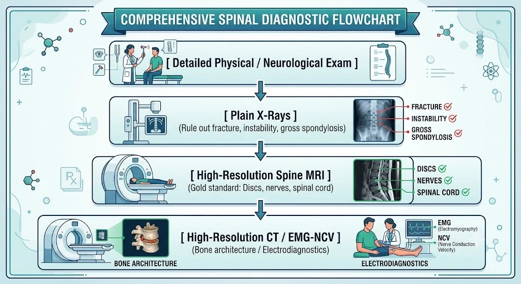
- Clinical Provocative Testing:
- Straight Leg Raise (SLR / Lasegue’s Sign): Elevation of the symptomatic leg between 30° and 70° reproducing true dermatomal pain below the knee indicates L4–S1 radiculopathy.
- Spurling’s Maneuver: Passive lateral neck flexion with axial compression that reproduces radiating upper extremity radicular pain indicates cervical nerve root entrapment.
- High-Field Magnetic Resonance Imaging (MRI): The gold standard for soft-tissue visualization. It defines disc hydration state, identifies radial annular tears, measures precise millimeter herniations (central, subarticular, or foraminal), and visualizes the degree of nerve root or spinal cord compression.
- Digital Radiography (X-Ray): Evaluates sagittal and coronal alignment, disc space narrowing, osteophytes, and dynamic stability using flexion-extension stress views to rule out spondylolisthesis.
- Electromyography & Nerve Conduction Studies (EMG/NCV): Delineates active nerve denervation from chronic peripheral neuropathies (e.g., diabetic neuropathy vs. true lumbar radiculopathy).
6. Comprehensive Multimodal Treatment Paradigm
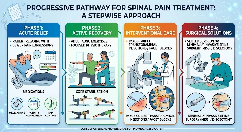
Ninety percent of patients suffering from an acute slipped disc, spondylosis, or back spasms recover without open surgery when treated using an evidence-based, multimodal conservative pathway.
Medical Pharmacotherapy
- NSAIDs: First-line treatment for chemical radiculitis and facet inflammation. COX-2 selective inhibitors (such as Etoricoxib or Celecoxib) or non-selective NSAIDs reduce prostaglandin cascade activation around the compressed nerve root.
- Skeletal Muscle Relaxants: Centrally acting agents (such as Thiocolchicoside, Chlorzoxazone, or Tizanidine) break the reflex pain-spasm-ischemia cycle in acute paraspinal contractures.
- Neuropathic Pain Modulators: Calcium channel alpha-2-delta ligands (Pregabalin, Gabapentin) stabilize hyper-excitable sensory neurons in radiating sciatica and brachialgia.
- Short-Course Oral Corticosteroids: High-potency methylprednisolone tapers may be considered in early acute disc herniations with profound radicular swelling, provided systemic contraindications are absent.
Advanced Interventional Pain Management
When systemic medications do not provide adequate relief, targeted image-guided interventions deliver high-potency anti-inflammatory agents directly to the point of nerve conflict:
- Transforaminal Epidural Steroid Injections (TFESI): Performed under C-arm fluoroscopic guidance. A micro-needle delivers a localized depot corticosteroid and local anesthetic directly into the neuroforamen adjacent to the herniated disc, resolving neurogenic edema.
- Facet Joint Injections & Radiofrequency Ablation (RFA): In cervical or lumbar spondylosis with localized facet arthrosis, fluoroscopy-guided injections into the articular capsule or thermal neurotomy of the medial branch nerves can eliminate mechanical back and neck pain.
7. Physiotherapy and Functional Spine Rehabilitation
Once the acute inflammatory phase stabilizes, an active exercise protocol is essential to stabilize the spinal columns, normalize mobility, and prevent chronic recurrence.
The McKenzie Method (Mechanical Diagnosis and Therapy – MDT)
The primary diagnostic framework for disc displacement focuses on directional preference:
- Centralization Phenomenon: The systematic performance of repetitive end-range movements (e.g., prone lumbar extensions) that cause distal radiating limb pain to migrate centrally toward the midline of the spine. Centralization correlates strongly with positive non-operative outcomes.
- Lumbar Extension Exercises: Prone press-ups gently leverage dynamic hydraulic pressures, guiding anterior movement of the displaced nucleus pulposus away from the dorsal nerve root.
Cervical Spine Mobilization & Isometric Strengthening
- Cervical Retraction (Chin Tucks): Realigns the cervical spine over the thoracic cage, reversing postural forward head tilt and reducing suboccipital tension.
- Multi-Planar Isometric Neck Strengthening: Gentle resistance against forward flexion, extension, and lateral bending without moving the joints, safely building deep neck flexor strength (longus colli, longus capitis).
- Scapular Stabilizer Training: Strengthening the middle and lower trapezius, rhomboids, and serratus anterior prevents thoracic kyphotic slump, which accelerates cervical disc degeneration.
Lumbo-Pelvic Core Stabilization
The lumbar spine relies on an internal muscular “corset” to neutralize axial loads:
- Deep Transverse Abdominis & Multifidus Activation: Recruited through abdominal drawing-in maneuvers (navel-to-spine) in supine or quadruped postures.
- The “Big Three” Stabilization Drills:
- Bird-Dog: Alternating contralateral arm and leg extensions in the quadruped position to train the posterior chain while maintaining a neutral spine.
- Side Plank: Activates the quadratus lumborum and lateral abdominal wall with minimal axial compressive loading on the discs.
- Modified Curl-Up: Activates the rectus abdominis while preserving natural lumbar lordosis, avoiding the intradiscal pressure spikes caused by traditional sit-ups.
- Pelvic Bridging: Restores gluteal activation and prevents hamstring overcompensation during hip extension.
8. Modern Surgical Innovations: Minimally Invasive Spine Surgery (MISS)
Surgical intervention is indicated when conservative management fails over a 6-to-12-week period, or immediately if progressive motor weakness, intractable radicular pain, or red flag symptoms appear.
Lumbar Microdiscectomy
The surgical benchmark for lumbar slipped discs. Performed through a 1.5–2 cm incision using a high-resolution operating microscope or high-definition endoscope:
- Subperiosteal paraspinal muscular dilation spares muscle tissue, preserving structural integrity.
- Targeted laminotomy and flavectomy expose the epidural space.
- The displaced disc fragment impinging on the traversing nerve root is excised, resolving root tension while preserving healthy disc tissue.
Anterior Cervical Discectomy and Fusion (ACDF) / Cervical Disc Arthroplasty (CDA)
- ACDF: For symptomatic cervical disc herniations and spondylotic radiculopathy/myelopathy. Approached through a transverse anterior neck crease, the entire disc and offending osteophytes are excised, decompressing the spinal cord and exiting nerve root. An interbody spacer (PEEK cage) packed with bone graft is inserted to re-establish disc height, backed by an anterior titanium stabilization plate.
- Cervical Disc Replacement (Arthroplasty): In young patients with isolated cervical disc herniations without severe facet arthritis, an artificial, motion-preserving disc prosthesis can be implanted to preserve normal kinetic motion and prevent accelerated adjacent segment disease.
9. Ergonomics, Prevention, and Spine Hygiene
Managing spine health requires active attention to daily biomechanics:
- Workstation Ergonomics: Set the computer monitor so the top third of the screen is at resting eye level. Keep elbows bent at 90 degrees with forearm support. Your feet should sit flat on the floor or on an angled footrest, with the lumbar curve supported by an adjustable chair cushion.
- The 30-Minute Break Rule: Prolonged static sitting doubles the intradiscal pressure compared to standing. Stand, walk, and perform light extension stretches every 30 minutes.
- Safe Lifting Biomechanics: Never bend at the waist with your knees straight. Keep heavy loads close to your chest, bend at the hips and knees, lock your core, and lift through your gluteal and quadricep muscles rather than pulling with your lumbar spine.
- Cervical Spine Screen Discipline: Avoid deep downward neck flexion while reading smartphones or tablets. Lift devices toward eye level to avoid generating high mechanical lever-arm forces on the C5–C7 cervical discs.
10. Frequently Asked Questions (FAQ)
Can a slipped disc heal on its own without surgery?
Yes. The majority of acute disc herniations regress through natural immune-mediated resorption. The body recognizes extruded nucleus pulposus in the epidural space as foreign tissue, triggering localized enzymatic breakdown and phagocytosis by macrophages. With rest, NSAIDs, and targeted physiotherapy, symptoms often resolve within 6 to 12 weeks.
How do I know if my pain is a muscle spasm or a slipped disc?
A paraspinal muscle spasm typically causes localized, severe aching and tightness across the back or neck, without pain traveling past the shoulder or knee. A slipped disc often triggers radiating radicular symptoms: electric shock-like sensations, sharp shooting pain, pins-and-needles paresthesia, or localized motor weakness along a specific dermatome in the arm or leg.
What is the most effective sleeping position for a slipped disc?
The best position maintains a neutral, unloaded spine:
Side Sleepers: Lie on your side with a supportive pillow keeping your neck level with your spine, and place a firm pillow between your knees to prevent pelvic and lumbar rotation.
Back Sleepers: Place a pillow underneath your knees (Fowler’s position). This relaxes the psoas muscle and reduces intradiscal pressure across the lumbar region.
Avoid Stomach Sleeping: Prone sleeping forces the cervical spine into maximum rotation and hyperextends the lumbar lordosis, increasing intradiscal and facet pressures.
When should I see a spine surgeon for sciatica or neck pain?
Seek specialist evaluation if your pain persists beyond 4 to 6 weeks despite conservative therapy, if pain radiates past your knee or elbow, or if you notice progressive limb weakness (such as catching your toes while walking or dropping items from your hands). Immediate emergency care is required if you experience loss of bowel or bladder control, numbness in your groin, or gait unsteadiness.
11. Expert Spine Care in Vadodara
Living with persistent back pain, neck stiffness, or radiating limb sciatica can limit your daily life. An accurate diagnosis and structured treatment plan can help you regain mobility and return to an active lifestyle.
At Ortho Care Hospital, we specialize in the comprehensive clinical diagnosis, non-operative rehabilitation, interventional pain care, and minimally invasive surgical management of cervical, thoracic, and lumbosacral spine conditions.
Lead Spine & Orthopedic Consultant
Owner, Medical Director, & Senior Orthopedic Surgeon
Ortho Care Hospital, Vadodara
Expertise: Advanced Trauma Care, Spine Clinic, Joint Replacement, Arthroscopy, and Sports Medicine.
Connect with Our Clinic
- Hospital Address: 2nd Floor, Sharnam Enclave, Above Kotak Bank, Subhanpura Main Road, New IPCL Road, Vadodara, Gujarat – 390023.
- Direct Appointment Phone: +91 093289 74527
- Official Web Portal: https://www.orthocarehospital.in/
- Google Business Profile & Clinic Directions: [suspicious link removed]
12. Clinical References & Evidence Base
- Deyo, R. A., & Mirza, S. K. (2016). Herniated Lumbar Intervertebral Disk. The New England Journal of Medicine, 374(18), 1763–1772.DOI: 10.1056/NEJMcp1512658
- Binder, A. I. (2007). Cervical Spondylosis and Neck Pain. BMJ (Clinical Research Ed.), 334(7592), 527–531.DOI: 10.1136/bmj.39127.608299.80
- Fardon, D. F., et al. (2014). Lumbar Disc Nomenclature: Version 2.0: Recommendations of the Combined Task Forces of the North American Spine Society, the American Society of Spine Radiology and the American Society of Neuroradiology. The Spine Journal, 14(11), 2525–2545.DOI: 10.1016/j.spinee.2014.04.022
- Weinstein, J. N., et al. (2006). Surgical vs Nonoperative Treatment for Lumbar Intervertebral Disc Herniation: The Spine Patient Outcomes Research Trial (SPORT): A Randomized Trial. JAMA, 296(20), 2441–2450.DOI: 10.1001/jama.296.20.2441
- McGill, S. M. (2010). Core Training: Evidence Translating to Better Performance and Injury Prevention. Strength and Conditioning Journal, 32(3), 33–46.Link: National Strength and Conditioning Association Resources
- Qaseem, A., et al. (2017). Noninvasive Treatments for Acute, Subacute, and Chronic Low Back Pain: A Clinical Practice Guideline from the American College of Physicians. Annals of Internal Medicine, 166(7), 514–530.DOI: 10.7326/M16-2367