1. Introduction: Knee Anatomy and the Cruciate Ligament Complex
The human knee joint is a complex hinge mechanism engineered to support body weight while accommodating multi-planar rotation, sudden deceleration, and pivoting. Joint stability relies heavily on four primary static stabilizers:
- Anterior Cruciate Ligament (ACL): Resists anterior translation of the tibia relative to the femur and controls rotational torque.
- Posterior Cruciate Ligament (PCL): Resists posterior translation of the tibia.
- Medial Collateral Ligament (MCL): Guards against valgus (inward) stress.
- Lateral Collateral Ligament (LCL) & Posterolateral Corner (PLC): Guard against varus (outward) stress and posterolateral rotation.
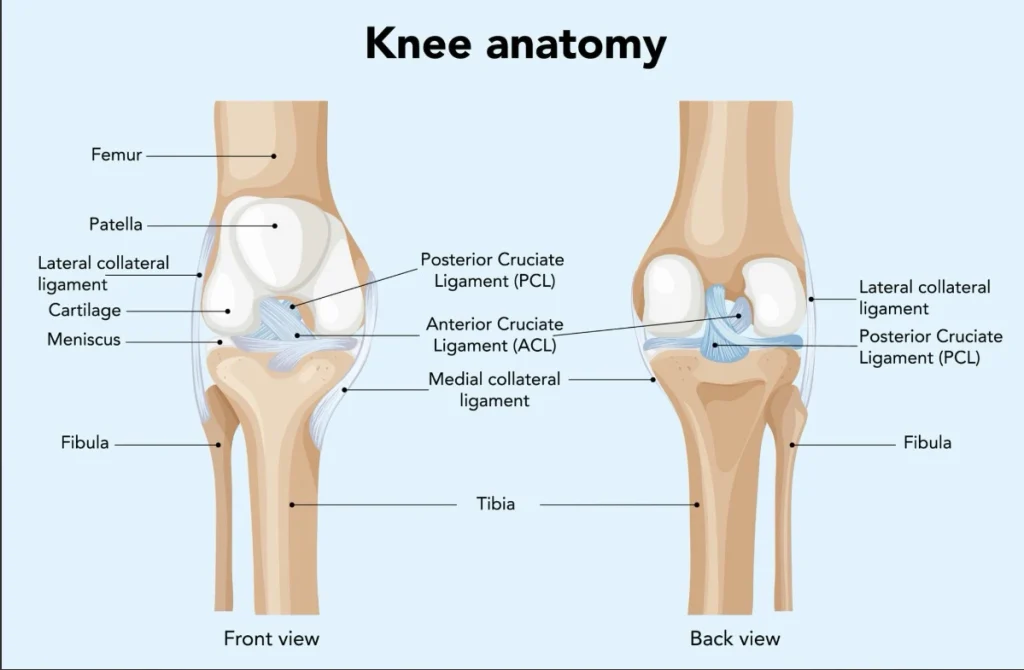
The ACL consists of two distinct functional bundles:
- Anteromedial (AM) Bundle: Tightest in knee flexion, controlling straight anterior translation.
- Posterolateral (PL) Bundle: Tightest near full extension, providing pivotal rotational stability.
When rotational or hyperextension force exceeds ligament tensile strength, an acl tear occurs. Modern arthroscopic interventions focus on anatomical cruciate ligament reconstruction, restoring native biomechanics through minimally invasive techniques that minimize surgical trauma and accelerate recovery.
2. Understanding Knee Ligament Tears: Partial vs. Full ACL Tear
An ACL injury typically occurs during non-contact sports maneuvers: sudden deceleration, jump landing on an extended knee, or rapid direction changes with the foot planted.
Grades of ACL Injury
- Grade 1 (Sprain/Micro-tear): Ligament fibers are stretched without mechanical elongation. The joint remains stable.
- Grade 2 (Partial Tear): Fibers are partially disrupted with mild laxity. Treatment depends on dynamic functional instability.
- Grade 3 (Full ACL Tear): Complete functional disruption of both bundles. The knee exhibits marked anterior and rotational instability. Because intra-articular synovial fluid washes away the blood clot necessary for native tissue repair, a complete tear cannot heal on its own.
Clinical Signs of an Acute Tear
- An audible or palpable “pop” at the moment of injury.
- Immediate, acute hemarthrosis (joint swelling with blood within 2–6 hours).
- Inability to continue physical activity or bear full weight.
- Persistent instability or an apprehension of the knee “giving way.”
Diagnostic Modalities
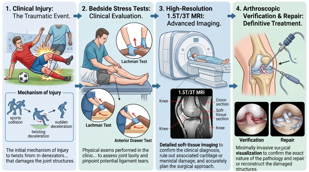
- Lachman Test: The most sensitive bedside physical examination test for anterior tibial displacement at 20°–30° of knee flexion.
- Pivot Shift Test: Evaluates dynamic rotational subluxation under valgus load; highly specific for functional instability.
- Magnetic Resonance Imaging (MRI): High-resolution imaging confirms tear status, evaluates graft donor sites, and detects associated injuries such as meniscal root tears, “bucket-handle” tears, or bone contusions (kissing contusions on the lateral femoral condyle and posterior lateral tibial plateau).
3. Indications & Criteria for Surgery: Conservative Management vs. Surgical Repair
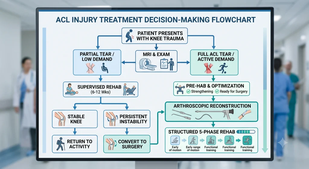
Not every ligament injury requires surgery. Modern ligament tear treatment is tailored to the patient’s physiological age, functional demands, occupational stresses, and degree of symptomatic instability.
| Parameter | Conservative Management Candidate | Surgical Reconstruction Candidate |
| Tear Degree | Grade 1 or low-grade partial tear | Full ACL tear or symptomatic partial tear |
| Activity Level | Sedentary lifestyle, low-impact activities (walking, cycling, swimming) | Competitive athletes, manual laborers, pivoting sports (football, cricket, badminton) |
| Knee Stability | Structurally stable on clinical stress testing | Evident rotational instability / positive Pivot Shift |
| Associated Pathologies | Isolated sprain without meniscal entrapment | Concomitant repairable meniscal tear or multiligament trauma |
| Risk of Secondary Damage | Low risk of cartilage wear | High risk of secondary osteoarthritis and meniscus degeneration if untreated |
4. Consulting a Specialized Sports Medicine Doctor
Evaluating complex joint injuries requires specialized training in joint preservation and kinetic biomechanics. A board-certified sports medicine doctor performs detailed clinical assessments to evaluate limb alignment, rotational deficits, and dynamic neuromuscular control.
At Ortho Care Hospital in Vadodara, under the clinical leadership of Dr. Sandeep Chauhan, arthroscopic surgery is guided by three principles:
- Preservation: Preserving healthy native tissue and meniscal volume whenever possible.
- Precision Placement: Placing anatomical tunnels at native footprints rather than non-anatomical vertical points.
- Holistic Recovery: Combining structural repair with objective functional testing to support safe return to full function.
5. Comprehensive Pre-Operative Planning & Graft Selection
Successful arthroscopic acl reconstruction depends on proper pre-operative preparation and individualized graft selection.
Pre-Habilitation (“Pre-Hab”)
Operating on an acutely inflamed, swollen knee with limited range of motion (ROM) increases the risk of post-operative arthrofibrosis (joint stiffness). Pre-hab focuses on:
- Resolving joint effusion using cryotherapy, compression, and anti-inflammatory therapy.
- Restoring full symmetric knee extension (0° neutral extension is mandatory before surgery).
- Reactivating quadriceps motor firing through isometric sets, straight leg raises, and cycling.
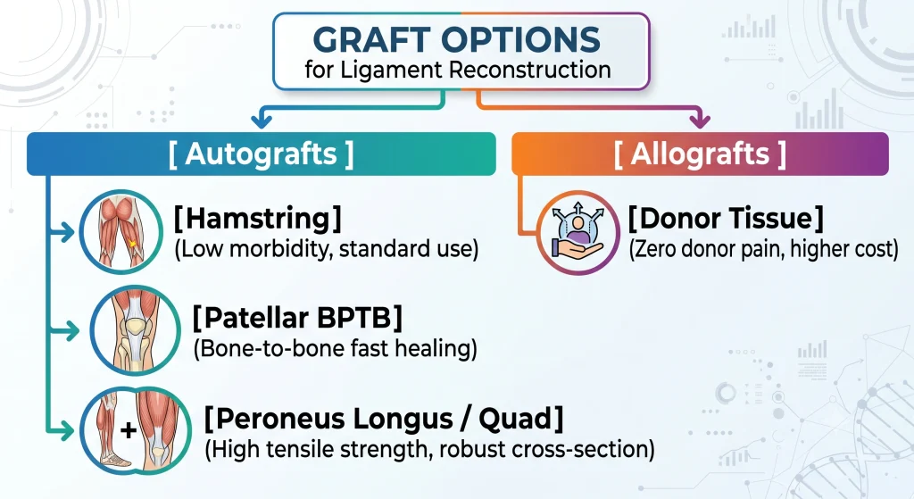
Graft Selection Matrix
Graft selection balances patient anatomy, sport-specific demands, revision considerations, and donor-site morbidity:
| Graft Type | Advantages | Disadvantages | Ideal Candidate |
| Hamstring Tendon (Quadrupled ST/G) | Minimal anterior knee pain; low incision morbidity; excellent tensile strength. | Tendon-to-bone healing takes longer; mild risk of residual hamstring weakness. | Recreational athletes, desk workers, general sports enthusiasts. |
| Bone-Patellar Tendon-Bone (BPTB) | “Gold Standard” for bone-to-bone healing (~6–8 weeks); rigid early fixation. | Higher incidence of anterior kneeling pain; small risk of patellar fracture. | High-level competitive pivoting athletes; revision surgeries. |
| Peroneus Longus Tendon | Excellent diameter (>8.5 mm without folding); avoids donor-site knee morbidity. | Requires ankle tendon harvesting; careful lateral ankle balance checks needed. | Multi-ligament reconstruction; patients with slim native hamstrings. |
| Quadriceps Tendon | Predictable graft diameter; minimal kneeling pain; balanced stiffness. | Harvesting requires specialized instrumentation; technically demanding. | Revision cases; young contact athletes. |
6. Step-by-Step Surgical Technique: Arthroscopic ACL Reconstruction
Modern arthroscopic reconstruction utilizes high-definition optical systems and specialized instrumentation to minimize surgical trauma.
The Surgical Procedure
- Diagnostic Arthroscopy: High-definition cameras inspect the patellofemoral joint, medial compartment, and lateral compartment. Accompanying meniscal tears are repaired or preserved (meniscectomy is kept to an absolute minimum).
- Graft Harvest & Preparation: The selected graft is harvested via a targeted incision, debrided of muscle tissue, whipstitched with non-absorbable sutures, pre-tensioned, and sized to 0.5 mm precision.
- Debridement & Notch Preparation: Remnants of the torn ACL are removed while preserving the tibial insertion footprint to retain native mechanoreceptors and proprioceptive nerve endings.
- Precision Tunnel Drilling:
- Femoral Tunnel: Drilled anatomically through an accessory anteromedial portal or via outside-in guides to replicate the native footprint on the lateral femoral condyle.
- Tibial Tunnel: Centered on the native tibial footprint, angled cleanly to avoid graft impingement against the intercondylar roof during full extension.
- Graft Passage & Tensioning: The graft is pulled into the joint through the tibial tunnel, docked securely into the femoral socket, and cycled through 20–30 full flexion-extension motions under tension to eliminate creep.
- Dual Fixation: The graft is secured using specialized femoral and tibial implants under controlled axial tension.
7. Choice of Implants & Fixation Hardware
Fixation devices provide mechanical stability while the graft incorporates into the bone tunnels.
Femoral Fixation
- Cortical Suspensory Buttons (Fixed Loop vs. Adjustable Loop):
- Mechanism: A low-profile titanium or PEEK button rests flat against the outer femoral cortex, suspending the graft within the tunnel.
- Modern Choice: Adjustable-loop buttons (e.g., UltraButton, TightRope) allow the surgeon to adjust tension precisely and dock graft tissue flush into the socket.
Tibial Fixation
- Bioabsorbable & Biocomposite Interference Screws:
- Materials: Poly-L-lactic acid (PLLA) combined with Tricalcium Phosphate (TCP) or Hydroxyapatite (HA).
- Benefit: Provides rigid compression within the tibial tunnel, gradually hydrolyzing over 3–5 years while stimulating bone ingrowth.
- PEEK (Polyether Ether Ketone) Screws:
- Non-absorbable, biocompatible polymer that maintains consistent strength indefinitely without MRI distortion.
- Secondary Backup Fixation: In high-demand cases or softer bone, surgeons often place a secondary low-profile cortical washer, staple, or screw post to secure the tibial suture limbs.
8. Cost of Surgery in Vadodara: Factors, Transparency, & Insurance
The cost of cruciate ligament reconstruction in Vadodara ranges between ₹75,000 and ₹1,95,000, depending on several clinical and logistical variables:
Key Cost Drivers
- Implant Choice: Premium US-FDA approved adjustable buttons, biocomposite screws, and meniscal repair devices (e.g., all-inside meniscal anchors) influence total material costs.
- Associated Pathologies: Treating an isolated ACL tear requires fewer surgical steps than managing a complex meniscus repair, chondral grafting, or secondary anterolateral ligament (ALL) reconstruction.
- Hospital Room Category: General ward, twin-sharing, or private deluxe accommodations adjust base bed charges, nursing expenses, and operative theater fees.
- Insurance & Cashless Mediclaim: Ortho Care Hospital partners with major Third-Party Administrators (TPAs), government health panels, and private health insurers for cashless hospitalization.
9. Post-Operative Rehabilitation Protocol: Phase-by-Phase Timeline
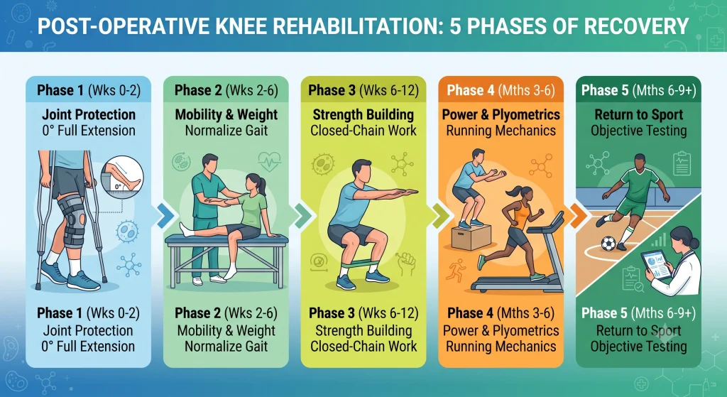
Successful outcomes require structured rehabilitation. The following timeline balances biological graft healing with functional progression:
Phase 1: Protection & Early Activation (Weeks 0–2)
- Goals: Protect graft fixation; resolve hemarthrosis; achieve full terminal knee extension (0°); restore active quadriceps firing.
- Weight Bearing: Toe-touch to partial weight-bearing with rigid hinged knee brace locked at 0° and axillary crutches.
- Exercises: Passive prone hangs (for extension), ankle pumps, patellar mobilizations, active-assisted knee flexion up to 90°, isometric quadriceps sets, straight leg raises (SLR) with no extension lag.
Phase 2: Mobility & Gait Normalization (Weeks 2–6)
- Goals: Progress knee flexion to 120°; achieve symmetrical full extension; transition off crutches to normal gait.
- Weight Bearing: Full weight-bearing as quadriceps control improves; unlock hinged brace past 30° once straight leg raises show no lag.
- Exercises: Stationary cycling with low resistance (initiating full revolutions), mini-squats (0°–45°), balance board training, hamstring curls, calf raises.
Phase 3: Strength & Neuromuscular Control (Weeks 6–12)
- Goals: Restore full knee range of motion; build functional kinetic chain strength (quads, hamstrings, glutes, core); improve single-leg balance.
- Discontinuation: Brace weaned off completely per surgeon guidance.
- Exercises: Leg press (up to 90°), Romanian deadlifts, step-ups and step-downs, lateral band walks, single-leg proprioceptive balance drills.
Phase 4: Power, Agility, & Plyometrics (Months 3–6)
- Goals: Build explosive strength; re-educate landing mechanics; begin straight-line running.
- Progression Criterion: Quadriceps index >75% compared to the contralateral leg; complete absence of joint effusion.
- Exercises: Linear treadmill running, ladder agility drills, double-leg landing mechanics transitioning to single-leg hops, eccentric loading exercises.
Phase 5: Sport-Specific Drills & Return-to-Sport Testing (Months 6–9+)
- Goals: Safe transition to pivoting, cutting, and full competitive athletic play.
- Objective Clearance Criteria (RTS Testing):
- Limb Symmetry Index (LSI) >90% on isokinetic strength tests for quadriceps and hamstrings.
- LSI >90% on functional single-leg hop tests (single hop, triple hop, crossover hop, timed 6-meter hop).
- Satisfactory dynamic valgus control during drop vertical jumps.
- Psychological readiness: High score on the ACL-Return to Sport after Injury (ACL-RSI) questionnaire.
10. Frequently Asked Questions (FAQ)
Can a full ACL tear heal on its own without surgery?
No. Because the anterior cruciate ligament is suspended in intra-articular synovial fluid, blood clots cannot form a stable biological scaffold across the gap. While non-operative rehabilitation can strengthen secondary stabilizers for low-demand activities, a full acl tear will not heal anatomically and often leads to recurring joint instability in active individuals.
What is the difference between an ACL repair and an ACL reconstruction?
An ACL repair stitches the patient’s torn ligament back onto the bone (suitable only for select proximal avulsion tears with high tissue quality, often reinforced with internal bracing). An arthroscopic acl reconstruction replaces the completely torn, degraded ligament with a biological tendon graft anchored into precisely drilled bone tunnels.
How soon can I return to driving after surgery?
Patients undergoing surgery on the left knee with an automatic vehicle can often drive within 2–3 weeks, once off narcotic medications. For right knee surgery or manual transmission vehicles, driving requires 6–8 weeks to ensure adequate braking response time and full quadriceps control.
Why is early pre-habilitation so important before surgery?
Undergoing surgery while the knee is stiff, swollen, and inflamed significantly increases the risk of post-operative arthrofibrosis (severe scar tissue formation). Pre-hab restores range of motion and baseline muscular control, leading to cleaner surgical visualization and faster post-operative rehab progression.
11. Schedule a Consultation at Ortho Care Hospital, Vadodara
If you are experiencing persistent knee instability, swelling, or pain following an athletic injury, an evaluation by an experienced specialist can help determine the right recovery path.
- Consultant Orthopaedic Surgeon: Dr. Sandeep Chauhan
- Hospital: Ortho Care Hospital
- Address: 2nd Floor, Sharnam Enclave, Above Kotak Bank, Subhanpura Main Road, New IPCL Road, Vadodara, Gujarat 390023
- Phone: 093289 74527
- Official Website: www.orthocarehospital.in
- Google Business Profile: Ortho Care Hospital on Google Maps
References
Dheerendra, S. K., Khan, W. S., Singhal, R., Shivarathre, D. G., Pydisetty, R., & Johnstone, D. (2012). Anterior Cruciate Ligament Graft Choices: A Review of Current Concepts. The Open Orthopaedics Journal, 6, 281–286. https://doi.org/10.2174/1874325001206010281 Cited by: 103
Frouin, A., Desfontaines, N., Lacourpaille, L., Nordez, A., & Le Sant, G. (2024). Preoperative Rehabilitation Enhances Mental and Physical Well-Being in Anterior Cruciate Ligament-Injured Individuals: A Mixed Methods Study. Journal of Sport Rehabilitation, 33, 289–296. https://doi.org/10.1123/jsr.2023-0312 Cited by: 4
Mouarbes, D., Menetrey, J., Marot, V., Courtot, L., Berard, E., & Cavaignac, E. (2019). Anterior Cruciate Ligament Reconstruction: A Systematic Review and Meta-analysis of Outcomes for Quadriceps Tendon Autograft Versus Bone–Patellar Tendon–Bone and Hamstring-Tendon Autografts. The American Journal of Sports Medicine, 47, 3531–3540. https://doi.org/10.1177/0363546518825340 Cited by: 592
Suomalainen, P., Kannus, P., & Järvelä, T. (2012). Double-bundle Anterior Cruciate Ligament reconstruction: a review of literature. International Orthopaedics, 37, 227–232. https://doi.org/10.1007/s00264-012-1680-9 Cited by: 55
Zarro, M., Dickman, M., Hulett, T., Rowland, R., Larkins, D., Taylor, J., & Nelson, C. (2023). Hop to It! The Relationship Between Hop Tests and The Anterior Cruciate Ligament – Return to Sport Index After Anterior Cruciate Ligament Reconstruction in NCAA Division 1 Collegiate Athletes. International Journal of Sports Physical Therapy, 18. https://doi.org/10.26603/001c.86130 Cited by: 14