
Persistent tingling in the fingers, nocturnal burning pain, and sudden loss of grip strength are classic presentations of Carpal Tunnel Syndrome (CTS). Left unaddressed, prolonged median nerve compression causes permanent sensory deficits and progressive atrophy of the thenar musculature.
Under the clinical direction of Dr. Sandeep Chauhan (Medical Director & Consultant Orthopaedic Surgeon), Ortho Care Hospital delivers comprehensive diagnosis, conservative management, and advanced open and endoscopic carpal tunnel release in Vadodara.
What Is Carpal Tunnel Syndrome?
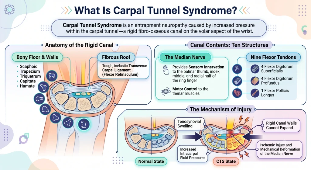
Carpal Tunnel Syndrome is an entrapment neuropathy caused by increased pressure within the carpal tunnel—a rigid fibro-osseous canal on the volar aspect of the wrist.
- Bony Floor & Walls: Formed by the carpal bones (scaphoid, trapezium, triquetrum, capitate, hamate).
- Fibrous Roof: Formed by the tough, inelastic transverse carpal ligament (flexor retinaculum).
- Canal Contents: Exactly ten structures traverse this rigid space:
- The median nerve (providing sensory innervation to the palmar thumb, index, middle, and radial half of the ring finger, plus motor control to the thenar muscles).
- Nine flexor tendons: 4 flexor digitorum superficialis, 4 flexor digitorum profundus, and 1 flexor pollicis longus.
Because the canal walls cannot expand, tenosynovial swelling or structural changes inevitably elevate intracarpal fluid pressures, causing ischemic injury and mechanical deformation of the median nerve.
Causes & Clinical Risk Factors
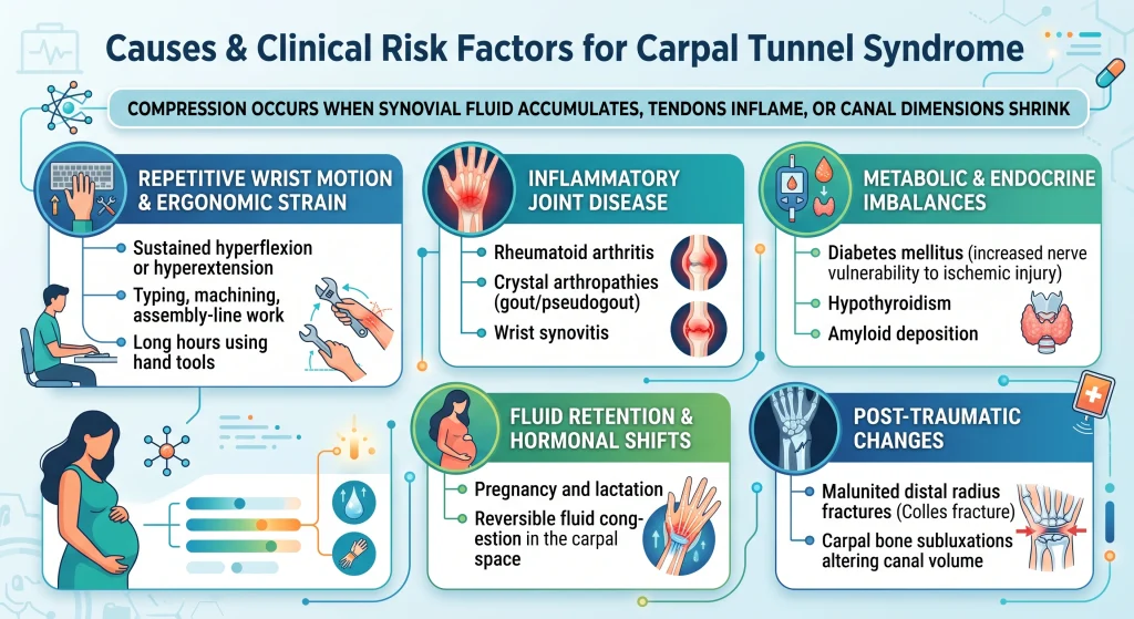
Compression occurs when synovial fluid accumulates, tendons inflame, or canal dimensions shrink:
- Repetitive Wrist Motion & Ergonomic Strain: Sustained hyperflexion or hyperextension during typing, machining, assembly-line work, and long hours using hand tools.
- Inflammatory Joint Disease: Rheumatoid arthritis, crystal arthropathies (gout/pseudogout), and wrist synovitis.
- Metabolic & Endocrine Imbalances: Diabetes mellitus (increased nerve vulnerability to ischemic injury), hypothyroidism, and amyloid deposition.
- Fluid Retention & Hormonal Shifts: Pregnancy and lactation frequently provoke reversible fluid congestion in the carpal space.
- Post-Traumatic Changes: Malunited distal radius fractures (Colles fracture) or carpal bone subluxations altering canal volume.
Signs and Symptoms
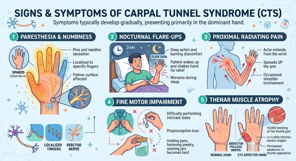
CTS symptoms typically develop gradually, presenting primarily in the dominant hand:
- Paresthesia & Numbness: “Pins and needles” sensation localized to the palmar surface of the thumb, index, middle finger, and the radial half of the ring finger. The little finger is spared (supplied by the ulnar nerve).
- Nocturnal Flare-ups: Patients routinely wake up with burning discomfort or deep aches, forced to “shake out” their hands (the Flick sign).
- Proximal Radiating Pain: Ache extending retrograde from the volar wrist into the forearm, elbow, and occasionally the shoulder.
- Fine Motor Impairment: Difficulty buttoning shirts, fastening jewelry, holding pens, or opening jars due to proprioceptive loss.
- Thenar Muscle Atrophy: In chronic, severe stages, visible wasting of the muscular pad at the base of the thumb (abductor pollicis brevis), leading to permanent weakness in thumb opposition.
Comprehensive Diagnostic Protocol at Ortho Care Hospital
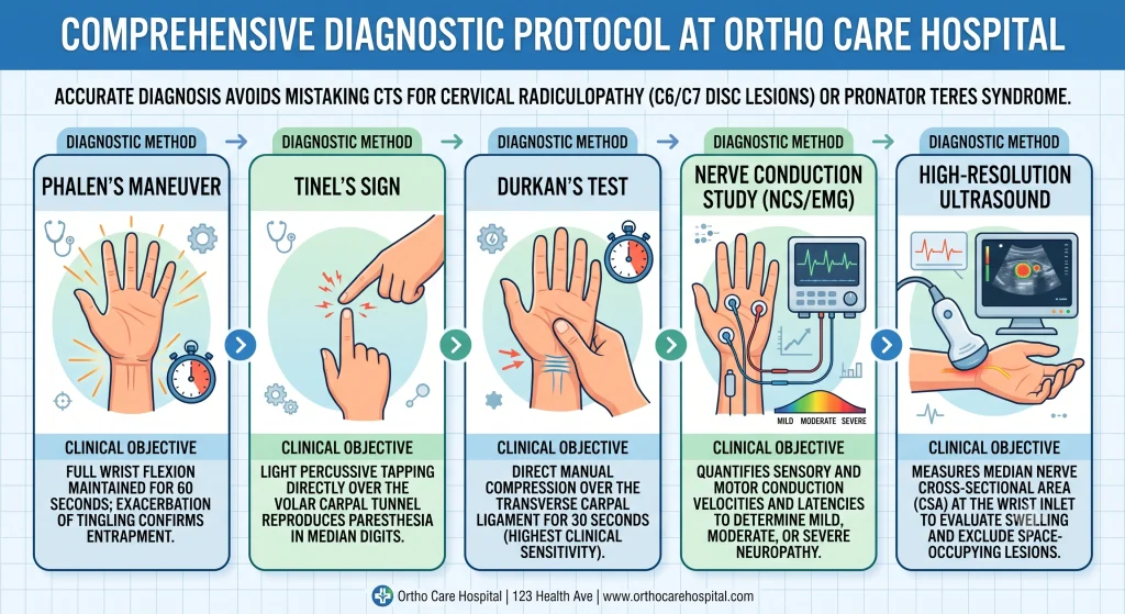
Accurate diagnosis avoids mistaking CTS for cervical radiculopathy (C6/C7 disc lesions) or pronator teres syndrome.
| Diagnostic Method | Clinical Objective |
| Phalen’s Maneuver | Full wrist flexion maintained for 60 seconds; exacerbation of tingling confirms entrapment. |
| Tinel’s Sign | Light percussive tapping directly over the volar carpal tunnel reproduces paresthesia in median digits. |
| Durkan’s Test | Direct manual compression over the transverse carpal ligament for 30 seconds (highest clinical sensitivity). |
| Nerve Conduction Study (NCS/EMG) | Quantifies sensory and motor conduction velocities and latencies to determine mild, moderate, or severe neuropathy. |
| High-Resolution Ultrasound | Measures median nerve cross-sectional area (CSA) at the wrist inlet to evaluate swelling and exclude space-occupying lesions. |
Stepwise Treatment Protocols
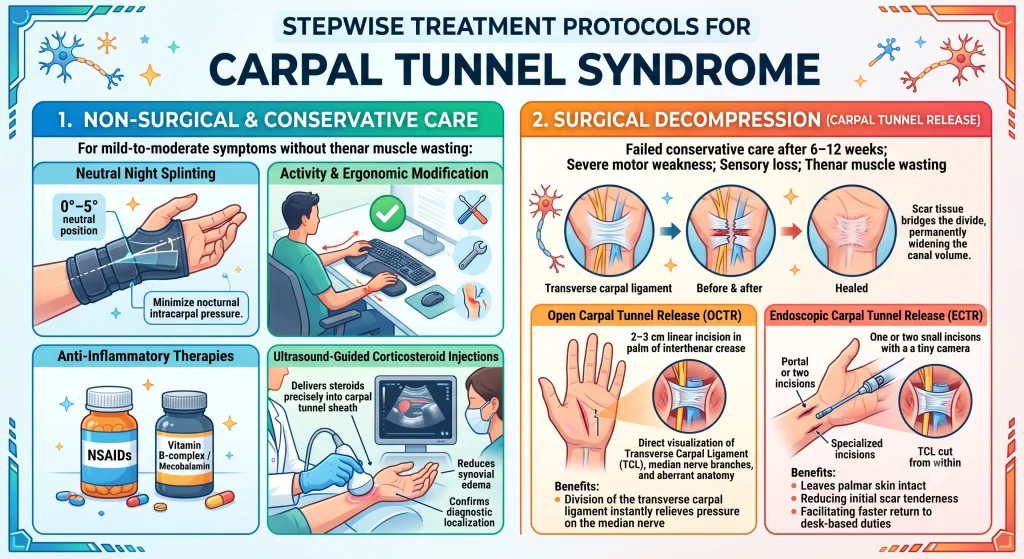
1. Non-Surgical & Conservative Care
For mild-to-moderate symptoms without thenar muscle wasting:
- Neutral Night Splinting: Cock-up wrist splints holding the joint in 0°–5° neutral position to minimize nocturnal intracarpal pressure.
- Activity & Ergonomic Modification: Repositioning desk equipment, keyboard rests, and mechanical tools to avoid sustained wrist flexion.
- Anti-Inflammatory Therapies: Targeted NSAIDs and neurotropic vitamin supplements (e.g., Vitamin B-complex / Mecobalamin).
- Ultrasound-Guided Corticosteroid Injections: Delivers anti-inflammatory steroids precisely into the carpal tunnel sheath, reducing synovial edema and confirming diagnostic localization.
2. Surgical Decompression (Carpal Tunnel Release)
Indicated when conservative care fails after 6–12 weeks, or in cases presenting with severe motor weakness, sensory loss, or thenar muscle wasting.
The procedure divides the transverse carpal ligament, instantly relieving pressure on the median nerve. As the ligament heals, scar tissue bridges the divide, permanently widening the canal volume.
- Open Carpal Tunnel Release (OCTR): A 2–3 cm linear incision is made in the palm along the interthenar crease. Allows direct, unobstructed visualization of the transverse carpal ligament, median nerve branches, and any aberrant anatomy.
- Endoscopic Carpal Tunnel Release (ECTR): Performed via one or two small incisions (portal technique) using a specialized cannula and camera. Leaves palmar skin intact, typically reducing initial scar tenderness and facilitating faster return to desk-based duties.
Post-Operative Rehabilitation & Timeline
- Days 1–3: Soft dressing applied; wrist elevated to reduce edema. Immediate active finger extension, flexion, and tendon-gliding exercises begin to prevent adhesions.
- Days 10–14: Suture removal. The wrist can be washed, and gentle scar massage commences with emollient creams.
- Weeks 2–6: Progressive range of motion and gradual grip-strengthening exercises. Avoid heavy lifting, power tool use, or forceful torque motions.
- Months 3–6: Complete scar remodeling. Chronic nerve regeneration proceeds at approximately 1 mm per day; long-standing sensory and motor deficits continue to improve over 6 to 12 months.
Why Choose Ortho Care Hospital Vadodara?
- Surgical Leadership: Evaluated and operated by Dr. Sandeep Chauhan, an experienced orthopaedic surgeon specializing in complex joint, trauma, and upper-limb reconstructive procedures.
- Precise Decompression: Microsurgical techniques that protect the delicate recurrent motor branch and palmar cutaneous branches of the median nerve.
- Integrated Care: On-site conservative regimens, splinting solutions, ultrasound-guided interventions, and dedicated post-operative hand physical therapy under one roof.
Frequently Asked Questions (FAQs)
Is carpal tunnel surgery performed under general anesthesia?
No. Most carpal tunnel release procedures are performed as outpatient day-care surgeries using local anesthesia or regional wrist/axillary nerve blocks. Patients remain awake and return home the same day.
How quickly does the tingling and pain disappear?
Nocturnal burning pain typically resolves within 24 to 48 hours following decompression. Numbness and sensory normalization take longer, depending directly on the severity and chronicity of pre-operative nerve compression.
Can carpal tunnel syndrome return after surgery?
True recurrence is rare (less than 2–5%). When persistent symptoms occur, they are typically related to incomplete release, perineural scarring, or secondary proximal nerve compression (double crush syndrome).
How do I know if my condition requires surgery instead of rest?
If you experience persistent daytime numbness, frequent object-dropping, or visible flattening of the muscle at the base of the thumb, surgery is typically indicated to prevent permanent nerve necrosis.
References
- American Academy of Orthopaedic Surgeons (AAOS). Management of Carpal Tunnel Syndrome Evidence-Based Clinical Practice Guideline.
- Atroshi, I., et al. “Prevalence of carpal tunnel syndrome in a general population.” JAMA, 282(2), 153-158.
- Bland, J. D. “Carpal tunnel syndrome.” BMJ, 335(7615), 343-346.
- Keith, M. W., et al. “Treatment of carpal tunnel syndrome.” Journal of the American Academy of Orthopaedic Surgeons, 17(6), 397-405.
Contact Ortho Care Hospital Vadodara
Schedule an in-person hand and wrist evaluation to evaluate your treatment options:
- Hospital Name: Ortho Care Hospital
- Director & Consultant: Dr. Sandeep Chauhan
- Address: 2nd Floor Sharnam Enclave, Above Kotak Bank, Subhanpura Main Road, New IPCL Road, Vadodara, Gujarat 390023
- Phone: 093289 74527
- Website: orthocarehospital.in